Cell Painting
Cell Painting is a high-content imaging technique that allows researchers to comprehensively profile and analyze the morphological characteristics of cells in response to different stimuli, treatments, or genetic perturbations. It provides a holistic view of cellular phenotypes, which can facilitate compound profiling, including mechanism of action elucidation and disease modelling. In addition, Cell Painting can be used in phenotypic drug discovery campaigns. The CBCS node at Uppsala University offers Cell Painting as a valuable service to collaborating researchers, enabling them to harness the power of this cutting-edge technology.
Labelling specific cellular components with fluorescent dyes or antibodies to create detailed morphological profiles. Commonly targeted structures include the nucleus, nucleoli, f-actin cytoskeleton, endoplasmic reticulum, Golgi apparatus, and mitochondria. The labelled cells are then imaged using high throughput/content microscopy, generating vast datasets of cellular images. The images are then analysed to generate rich-data that is used for profiling or drug discovery purposes.
Representative image of Human osteosarcoma U-2 OS cells stained using the Cell Painting protocol. For simplicity, in this image only 3 out of the 6 stains are visualised, that is Hoechst 33342 for the Nucleus, Mitotracker for the Mitochondria and Phalloidin+Wheat Germ Agglutinin for the F-Actin cytoskeleton and Golgi apparatus.
Functional Precision Medicine
CBCS supports researchers in conducting studies related to Functional Precision Medicine. This approach involves profiling disease models against libraries of compounds to identify and characterize their functional effects. By combining complex disease models, such as patient-derived cells or genetically engineered models, with high-throughput screening, researchers can gain insights into personalized treatment strategies and identify compounds with specific therapeutic potential.
The current services in functional precision medicine include:
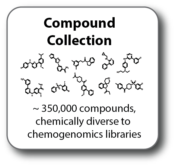
Drug library design and delivery
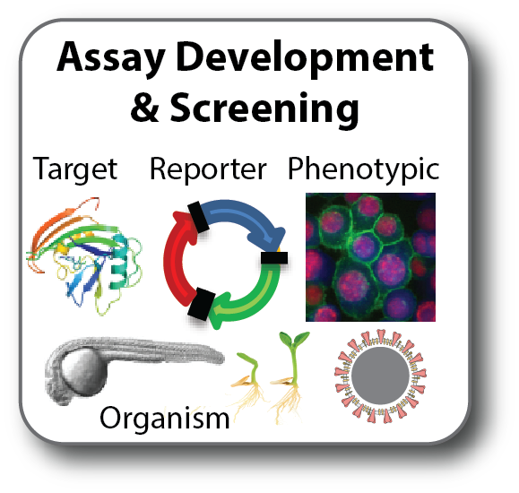
Assay development
Guidance and support regarding sample logistics, and best practices
Electrophysiology - understanding small molecule effects on ion channels.
Dr. Nina Ottosson at The CBCS Linköping electrophysiology unit, operating the automated patch clamp QPatch 48X.
Electrophysiology encompasses the investigation of proteins pivotal for the electrical signaling within the body, enabling vital processes such as cardiac rhythm regulation and neural transmission. These proteins, commonly referred to as ‘ion channels’, are embedded within membranes where they orchestrate the flux of ions across membrane barriers, such as cell a membrane.
At the electrophysiology node of CBCS at Linköping University, we use the automated patch clamp (APC) technique to investigate the functions and properties of ion channels comprehensively. This technique spans the entire spectrum of applications, ranging from detailed academic ion channel characterization to primary screening for new ion channel targeting drugs. We are happy to help you with your research, regardless of whether you are an experienced electrophysiologist or if you are new to electrophysiology.
Our support encompasses diverse applications, including but not limited to the following examples:
Basic Research and Characterization of Ion Channels
Ion Channel Mutations
Drug Discovery for ion channel targeted drugs
Safety pharmacology
Learn more about the services offered through collaboration with the CBCS or contact us directly to discuss your research needs.


